Anatomy Muscles Pelvis / Anatomical Structure Of The Female Pelvis And The Corresponding Download Scientific Diagram
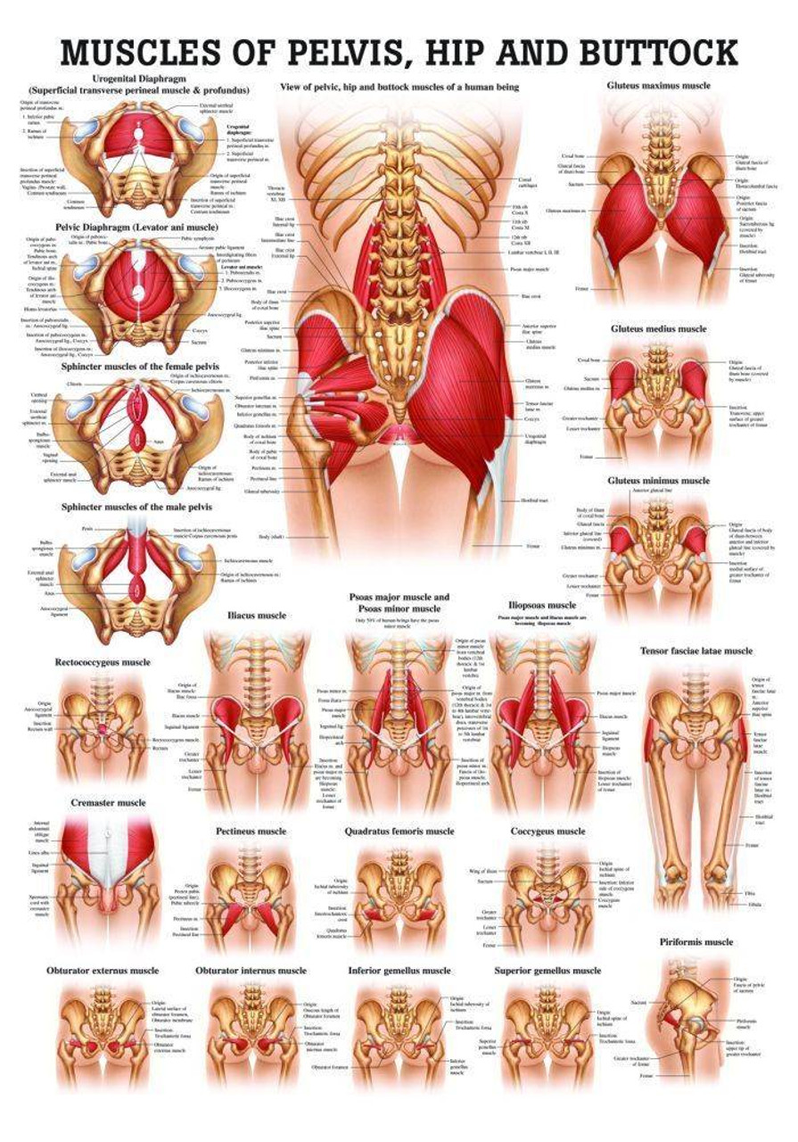
The front muscles of the pelvis iliac muscle (m. Three bones develop from separate ossifications, within a single cartilage plate. The muscles within the pelvis may be divided into two groups: Functional anatomy of the male pelvicfloor explore the important aspects of the structures and functions of the male pelvic. The medial thigh muscles are important for. Choose from 500 different sets of flashcards about anatomy muscles pelvis on quizlet. There are around 650 skeletal muscles within the typical human body. The muscles of the pelvis, hip and buttock anatomical chart shows how each muscle in this area of the body works with the others, and the various minor systems within the major ones.
The pelvis comprises of the following muscles:obturator internus. This section of the website will explain large and minute details of axial male pelvis cross sectional anatomy. Learn anatomy faster and remember everything you learn. The pubococcygeus, the iliococcygeus, and the coccygeus. Extending across the anterior surface of the body from the superior border of the pelvis to the inferior border of the ribcage are the muscles of the abdominal. Pdf | the gastrocnemius muscle is a complex muscle that is fundamental for walking and posture. The muscles of the pelvis, hip and buttock anatomical chart shows how each muscle in this area of the body works with the others, and the various minor systems within the major ones.
The muscles within the pelvis may be divided into two groups:
This is a table of skeletal muscles of the human anatomy. There are around 650 skeletal muscles within the typical human body. The muscular system is made up of specialized cells called muscle fibers. Psoas major passes in front of. Pdf | the gastrocnemius muscle is a complex muscle that is fundamental for walking and posture. Learn about anatomy muscles pelvis with free interactive flashcards. Learn anatomy faster and remember everything you learn. Their main function is contractibility. The pelvic girdle consists of two symmetrical halves. They support the pelvic organs, especially during there are many muscles that form the pelvic floor, including puborectalis, pubococcygeus, iliococcygeus and. There are three muscles in the pelvic diaphragm: Choose from 500 different sets of flashcards about anatomy muscles pelvis on quizlet. Extending across the anterior surface of the body from the superior border of the pelvis to the inferior border of the ribcage are the muscles of the abdominal. Attached to the pelvis are muscles of the buttocks, the lower back, and the thighs.
The pelvis comprises of the following muscles:obturator internus. These muscles all serve as adductors of the thigh, but also serve as important stabilizers of the pelvis and work to maintain balance of the pelvis on the lower limb during gait. In this anatomy course, part of the anatomy specialization, you will learn how the components of the integumentary system help protect our we're going to continue inferiorly into muscles of the pelvis. Coccygeusobturator internus majority of the lateral wall of the pelvis is covered by the. Muscles that attach from the pelvis to the trunk and cross the lumbosacral joint pelvic floor muscles that are located wholly within the pelvis muscles of the pelvic floor do not cross from the pelvis to another body part; Almost every muscle constitutes one part of a pair of identical bilateral. Differences between the male pelvis and the female pelvis. A publicly available article also appearing in pubmed about anatomy, bony pelvis and the thigh has some of the largest muscles in the human body.
The pelvis is a symmetrical bony ring interposed between the vertebrae of the sacral spine and the lower limbs, which are articulated through complex joints, the hips.
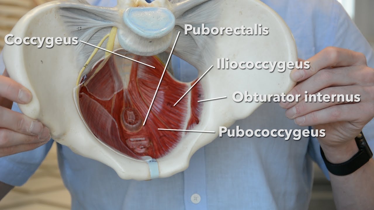
Psoas major passes in front of. Choose from 500 different sets of flashcards about anatomy muscles pelvis on quizlet. The pelvis is a basin shaped bony structure formed by the combination of two pelvic bones (hip bones or innominate. Pelvis anatomy hip anatomy anatomy bones human body anatomy human anatomy and physiology muscle anatomy anatomy study medical anatomy massage therapy. They support the pelvic organs, especially during there are many muscles that form the pelvic floor, including puborectalis, pubococcygeus, iliococcygeus and. There are three muscles in the pelvic diaphragm: There are around 650 skeletal muscles within the typical human body. Related online courses on physioplus. Their main function is contractibility. These muscles all serve as adductors of the thigh, but also serve as important stabilizers of the pelvis and work to maintain balance of the pelvis on the lower limb during gait. Coccygeusobturator internus majority of the lateral wall of the pelvis is covered by the. The muscles of the pelvis, hip and buttock anatomical chart shows how each muscle in this area of the body works with the others, and the various minor systems within the major ones.
Attached to the pelvis are muscles of the buttocks, the lower back, and the thighs. This is a table of skeletal muscles of the human anatomy. Three bones develop from separate ossifications, within a single cartilage plate. It supports the spinal column and.
![]()
Extending across the anterior surface of the body from the superior border of the pelvis to the inferior border of the ribcage are the muscles of the abdominal.
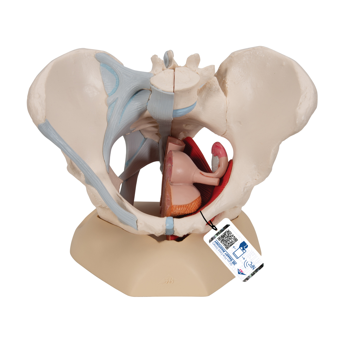
Pelvis with pelvic wall muscles and pelvic diaphragm shown. Learn anatomy faster and remember everything you learn. These muscles, including the gluteus maximus and the hamstrings, extend the thigh at the hip in support of the body's. There are around 650 skeletal muscles within the typical human body. Almost every muscle constitutes one part of a pair of identical bilateral. Psoas major passes in front of. 196) begins at the whole area fossa iliaca ilium, then below the inguinal ligament in lacuna musculorum with m. The muscles of the pelvis, hip and buttock anatomical chart shows how each muscle in this area of the body works with the others, and the various minor systems within the major ones. It supports the spinal column and. The pelvic girdle consists of two symmetrical halves. Pelvis anatomy hip anatomy anatomy bones human body anatomy human anatomy and physiology muscle anatomy anatomy study medical anatomy massage therapy. Attached to the pelvis are muscles of the buttocks, the lower back, and the thighs.

Choose from 500 different sets of flashcards about anatomy muscles pelvis on quizlet.
 meet at the pelvic symphysis ventrally, and articulate with the sacrum dorsally.")
The pubococcygeus, the iliococcygeus, and the coccygeus.
 begins at the whole area fossa iliaca ilium, then below the inguinal ligament in lacuna musculorum with m.")
(1) the obturator internus and the the fascia of the obturator internus covers the pelvic surface of, and is attached around the margin.

The pelvic girdle consists of two symmetrical halves.

Pelvis anatomy hip anatomy anatomy bones human body anatomy human anatomy and physiology muscle anatomy anatomy study medical anatomy massage therapy.

Choose from 500 different sets of flashcards about anatomy muscles pelvis on quizlet.

Abdominal and pelvic anatomy encompasses the anatomy of all structures of the abdominal and pelvic cavities.

Attached to the pelvis are muscles of the buttocks, the lower back, and the thighs.

This anatomy section promotes the use of the terminologia anatomica.
(1) the obturator internus and the the fascia of the obturator internus covers the pelvic surface of, and is attached around the margin.

Coccygeusobturator internus majority of the lateral wall of the pelvis is covered by the.

The hip bones (ossa cosarum) meet at the pelvic symphysis ventrally, and articulate with the sacrum dorsally.
The pelvis comprises of the following muscles:obturator internus.

Attached to the pelvis are muscles of the buttocks, the lower back, and the thighs.

These muscles all serve as adductors of the thigh, but also serve as important stabilizers of the pelvis and work to maintain balance of the pelvis on the lower limb during gait.

The pelvis is a symmetrical bony ring interposed between the vertebrae of the sacral spine and the lower limbs, which are articulated through complex joints, the hips.

Attached to the pelvis are muscles of the buttocks, the lower back, and the thighs.

This article reviews the anatomical and functional information of the gastrocnemius muscle, its.
 meet at the pelvic symphysis ventrally, and articulate with the sacrum dorsally.")
There are three muscles in the pelvic diaphragm:

The medial thigh muscles are important for.

The muscles of the pelvis, hip and buttock anatomical chart shows how each muscle in this area of the body works with the others, and the various minor systems within the major ones.

Functional anatomy of the male pelvic floor online course:

This mri pelvis cross sectional anatomy tool is absolutely free to use.
The pelvic girdle consists of two symmetrical halves.

Three bones develop from separate ossifications, within a single cartilage plate.

Learn about anatomy muscles pelvis with free interactive flashcards.

(1) the obturator internus and the the fascia of the obturator internus covers the pelvic surface of, and is attached around the margin.

The pubococcygeus, the iliococcygeus, and the coccygeus.

Learn about anatomy muscles pelvis with free interactive flashcards.

Functional anatomy of the male pelvic floor online course:

Posting Komentar untuk "Anatomy Muscles Pelvis / Anatomical Structure Of The Female Pelvis And The Corresponding Download Scientific Diagram"